

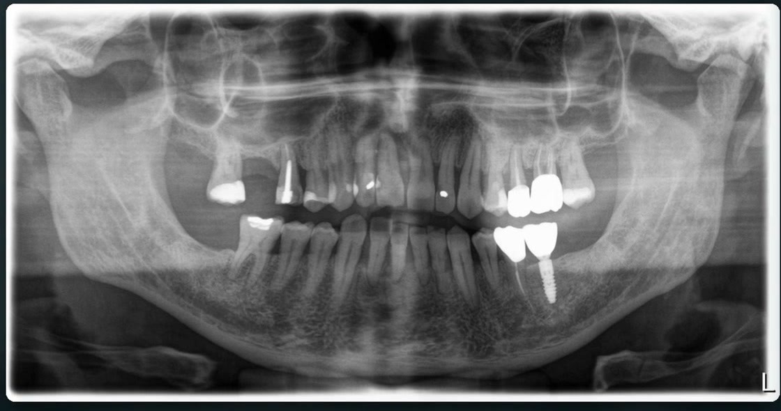
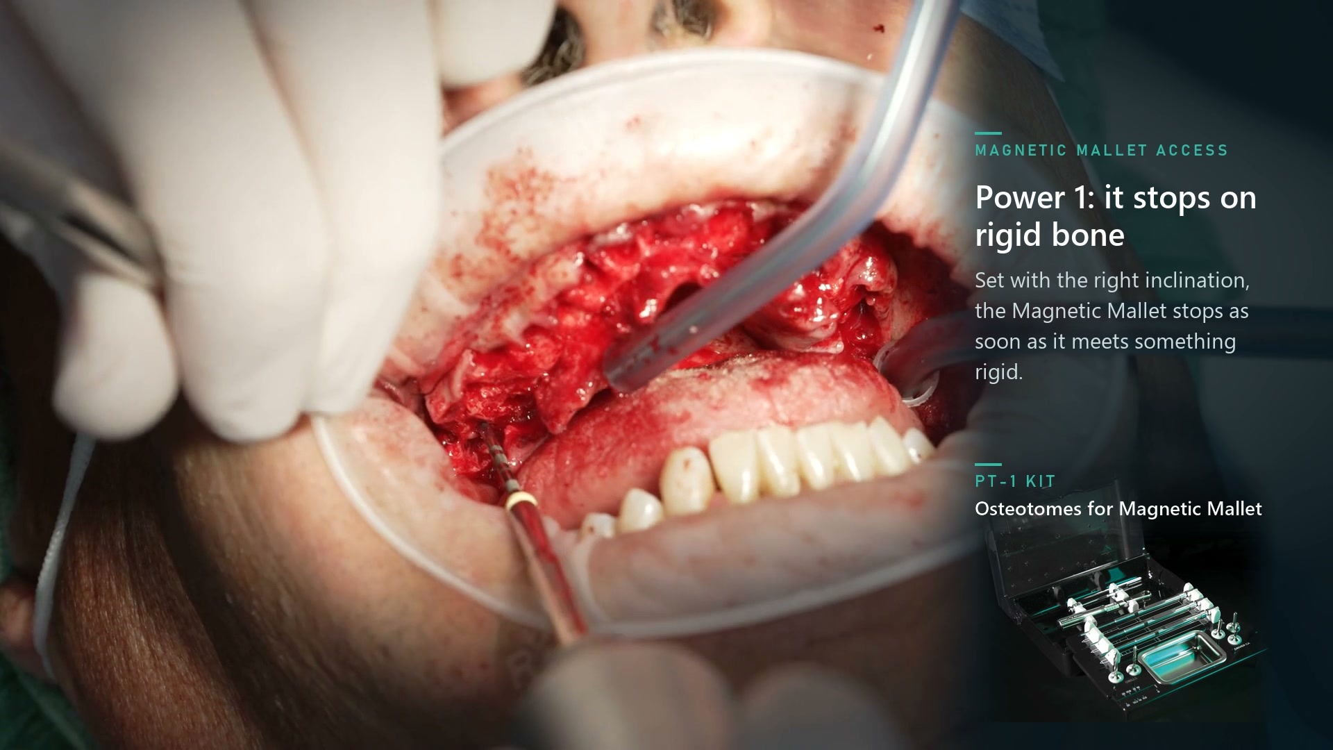
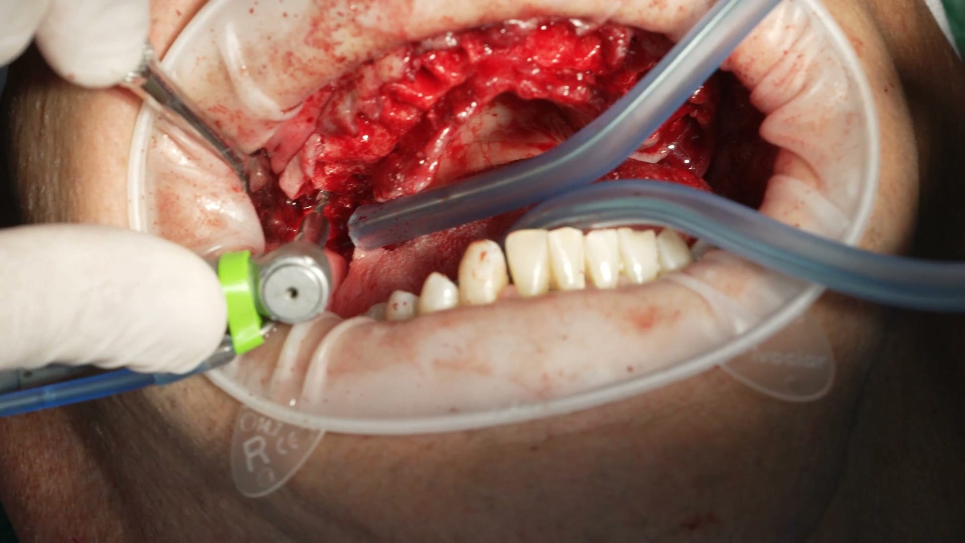
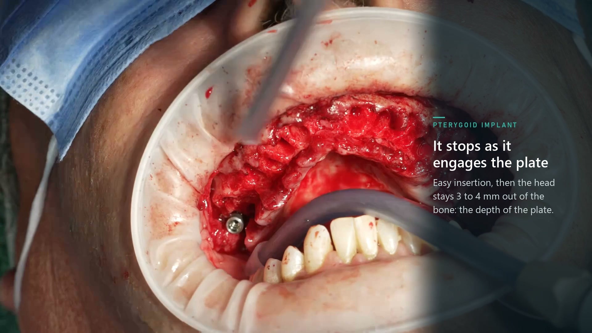
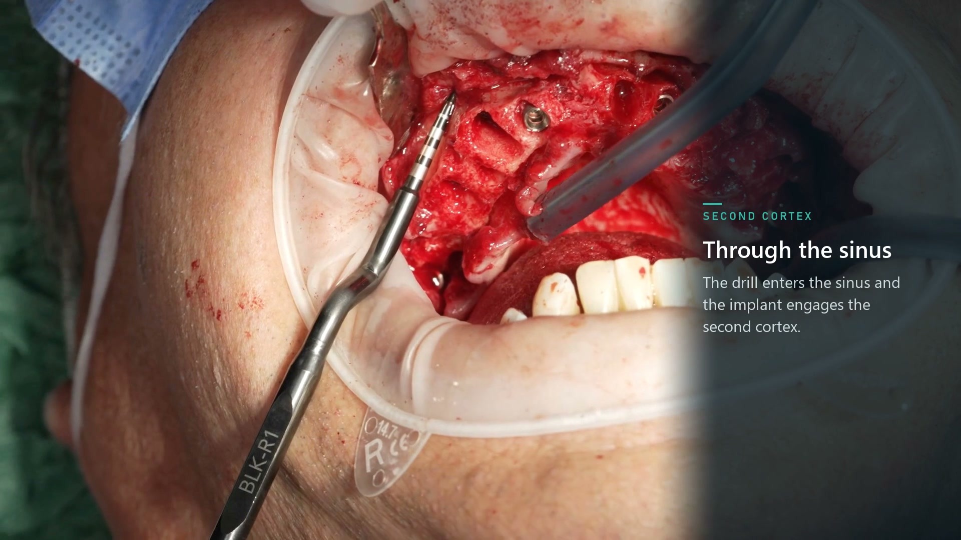
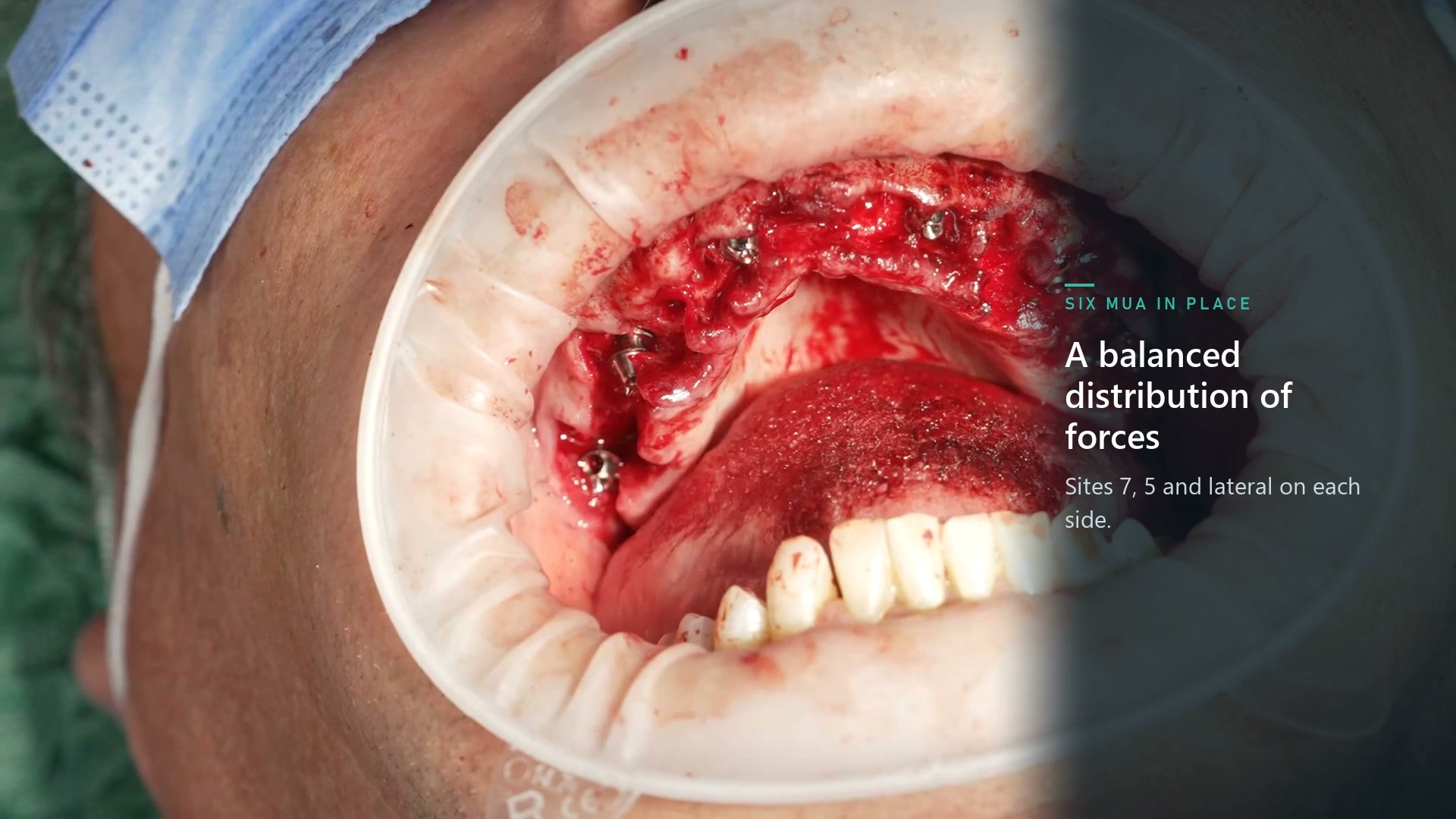
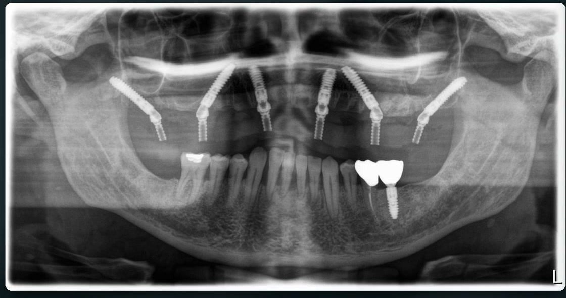
Clinical case of 9 November 2026 · Speaker Dr. Andrea Malavasi In a low-density upper jaw, where should anchorage be sought for the six implants, and how should their inclination be managed with the prosthesis in mind? Dr. Andrea Malavasi presents a case with two pterygoid implants and four anterior implants, two in the lateral incisor region and two in the second premolar region. Preparation of the pterygoid sites. After raising a full-thickness flap, Malavasi regularizes the ridge and locates the posterior access point with the help of a probe. In this case he interprets the stop of the Magnetic Mallet, set to power 1, as reaching the pterygoid plate. He records the depth with a rubber stop, completes the preparation with the PT-1 kit and passes through the plate with a drill. He repeats the sequence on the opposite side. Anterior anchorage and trans-sinus sites. The four anterior implants seek anchorage in the cortical bone of the nasal floor. In the sites with a trans-sinus path, Malavasi shows how he uses osteotomes to compact the first millimetres of crestal bone. The drill then crosses the sinus section and the implant engages the second cortex. Prosthetic choices and final check. The speaker shows how he orients the multi-unit abutments (MUAs) according to the emergence of the screws and the space to the opposing arch. On the lateral implants he uses two 17° MUAs to correct the bucco-palatal inclination. After placing the six MUAs, he checks the fit with the transfers in place and takes the impression. The postoperative panoramic shows the distribution of the six implants. Case images 1. Preoperative panoramic. Initial radiographic situation of the upper jaw. 2. Posterior access with the Magnetic Mallet. Orientation of the instrument along the pterygoid trajectory. 3. Preparation of the plate with the drill. Switching from the osteotome to the drill to pass through the cortex. 4. Insertion of the pterygoid implant. Position of the implant as it begins to engage the plate, before final tightening. 5. Preparation of the trans-sinus site. Advancing towards the second cortex after compaction of the crestal bone. 6. Six MUAs in place. Intraoperative view of the distribution of the abutments along the arch. 7. Postoperative panoramic. Final result with the two pterygoid implants and the four anterior implants. Question for colleagues When the axis of a pterygoid implant leads to a very vertical emergence, what reference do you use to choose the MUA angulation and keep space to the opposing arch?
Assisted translation
Read the original conversation